Aperture · DICOM viewer
What Aperture can do
Aperture opens CT, MR and radiography studies in the browser: slices, 3D reconstruction, 4D time series, measurements, and an AI assistant that can drive the viewer. Every screenshot below comes from a public scan you can open yourself after signing in: the built-in examples, and for the abdominal CT screen, a scan linked in that section.
Aperture is for research use only. It is not a medical device and must not be used for diagnosis, treatment or any clinical decision.
Open a study
Choose Import study or drop files anywhere on the page. Aperture reads single DICOM files (extensions are optional), whole folders including subfolders, and ZIP archives. Nothing is uploaded to open a study: files are parsed on your device and saved to your library. Import results list anything that was skipped and why.
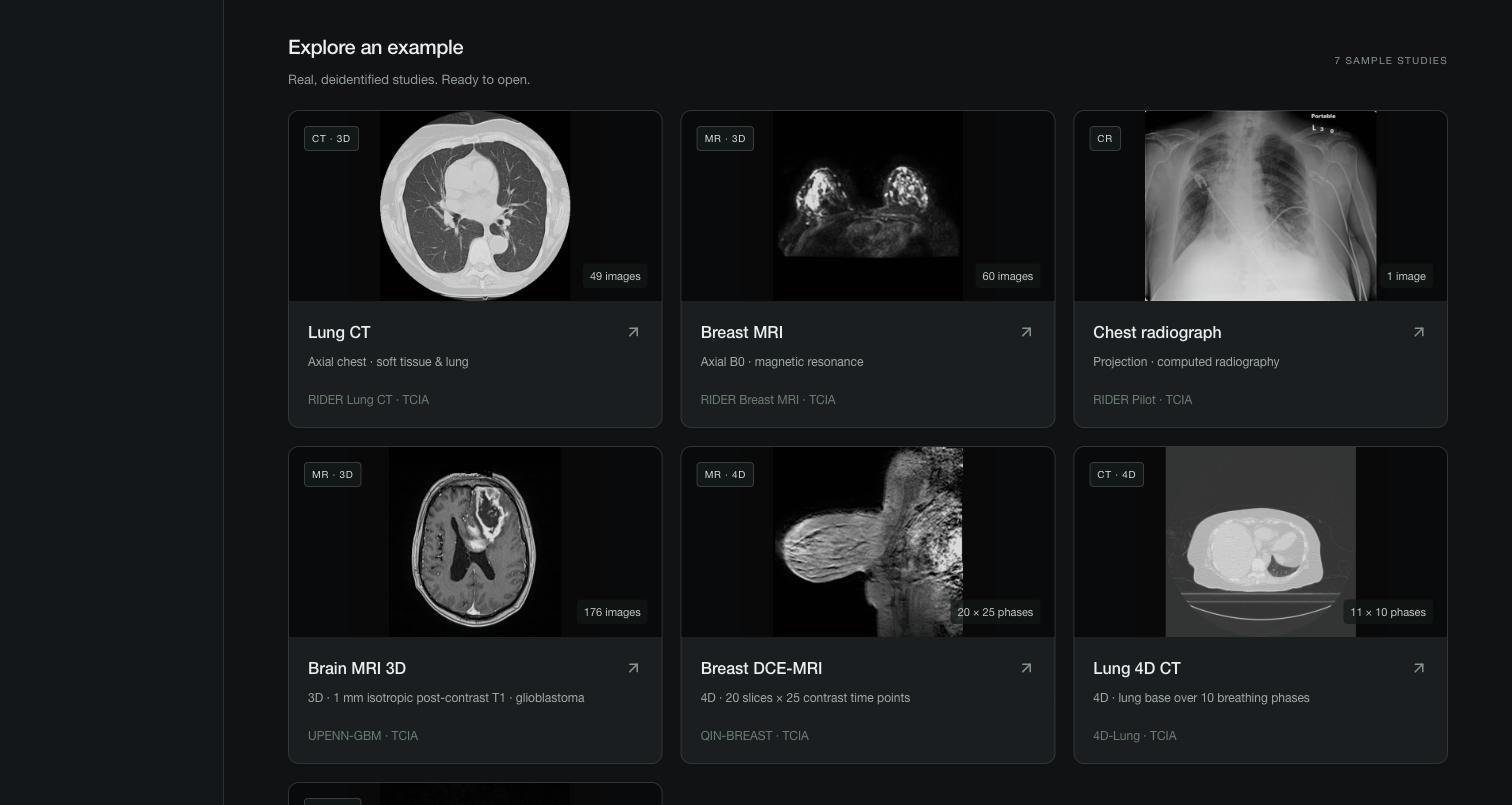
Or open one of the seven examples, all real, deidentified scans from The Cancer Imaging Archive. Their cards say which ones are 3D volumes and which are 4D.
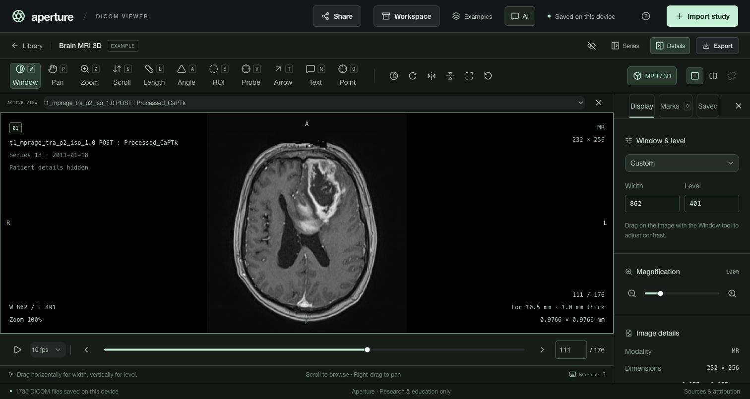
Open Examples → Brain MRI 3D. It is 176 slices of a post-contrast brain MRI at 1 mm, with a glioblastoma in the left frontal lobe.
View and navigate
- Scroll with the wheel, the arrow keys or the slider. Home / End jump to the first or last image, and PgUp / PgDn move ten at a time. Type a number in the box to jump to that image.
- Window / level: drag with the Window tool (W) or type width and level. CT presets: 1 lung (1500 / −600), 2 soft tissue (400 / 40), 3 bone (1800 / 400), 4 brain (80 / 40), 5 abdomen (350 / 50). 0 restores the DICOM window and 6 sets auto contrast.
- Zoom (Z, middle-drag), pan (P, right-drag), rotate, flip, invert (I), fit (F) and reset (R).
- Cine plays at 1–30 fps with Space.
- The overlay shows orientation letters derived from the patient geometry, window, zoom, slice location and thickness, and pixel spacing. Patient details stay hidden until you choose to show them.
Measure and mark
Measurement tools use the DICOM pixel spacing, so lengths are in millimetres, and CT intensities are in Hounsfield units when the rescale values allow it. Without spacing, Aperture says so and measures in pixels.
- L length, A angle, E ellipse ROI (area, mean, SD), V probe (one value).
- T arrow, N text label, Q numbered point.
- The Marks tab lists every mark in the study. You can rename, recolor, hide or delete each one, and undo or redo changes. Clicking a mark opens the exact series and slice it was drawn on. H hides all marks while you read the image.
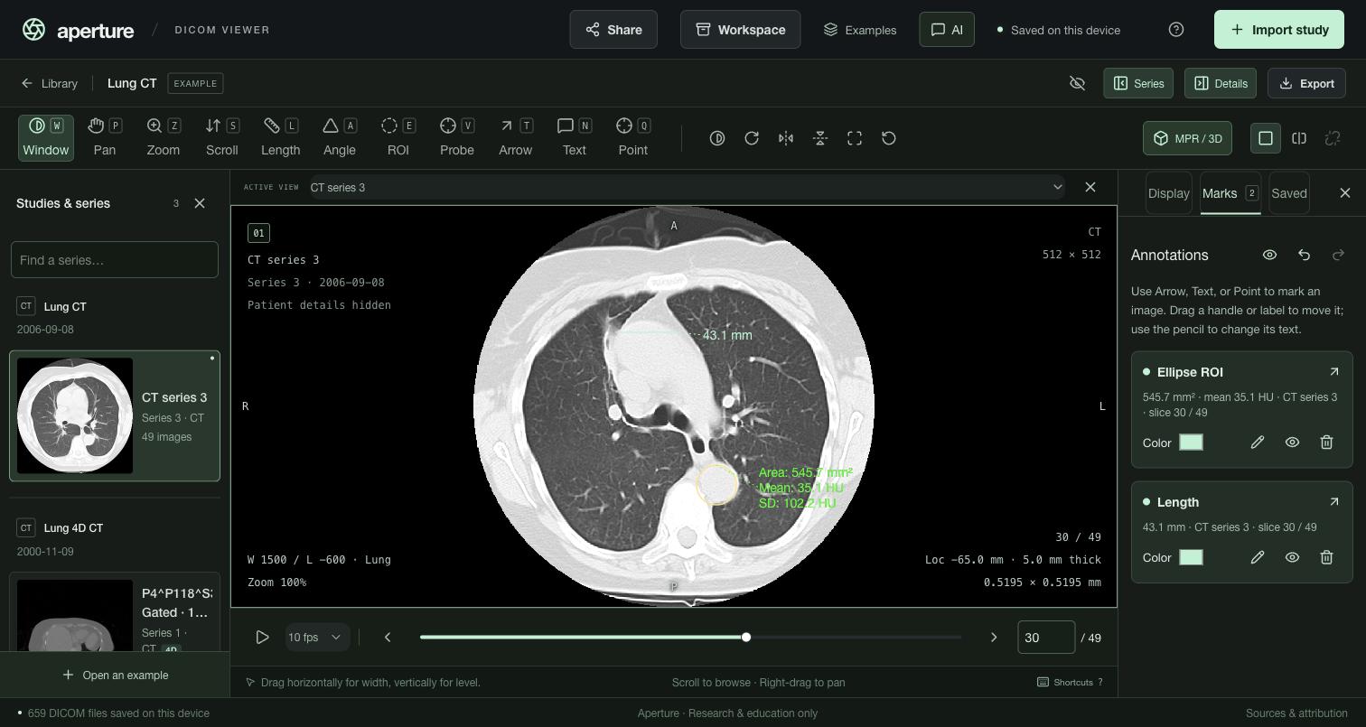
Open Lung CT, go to image 30, press 1, then L, and drag across the aorta.
Compare two series
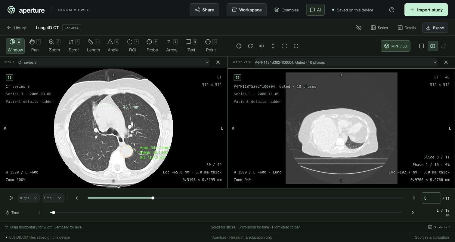
The two-view layout puts any two series side by side, from the same study or different ones. Each view has its own series picker and remembers its display. When both series share a frame of reference and orientation, Link keeps them on the nearest physical slice as you scroll. Two linked 4D series with the same number of phases also stay in step through time.
MPR and 3D
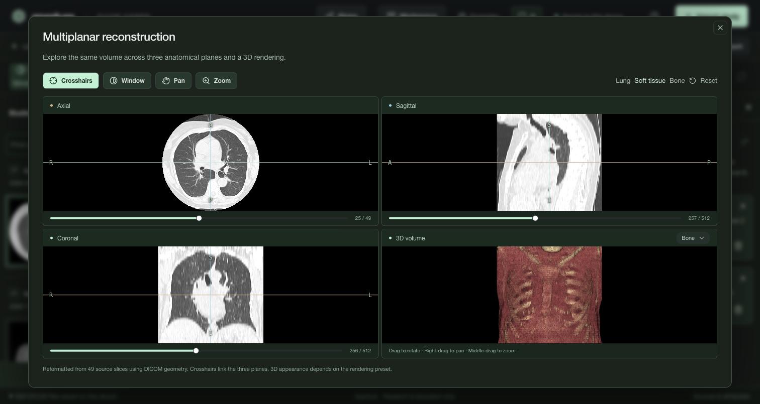
MPR / 3D rebuilds a regular CT or MR stack as a volume. It shows axial, sagittal and coronal planes linked by crosshairs, each with a slider, plus a rotatable 3D rendering with presets. CT gets lung, soft-tissue and bone windows. Aperture checks the geometry first: tilted, unevenly spaced, repeated or mixed slices are refused, with the reason in the button's tooltip.
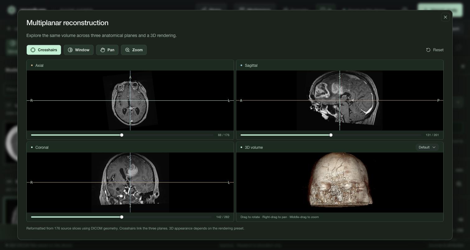
Open Brain MRI 3D, then MPR / 3D. Drag the crosshairs onto the tumor, and drag the 3D view to rotate it.
4D: slices over time
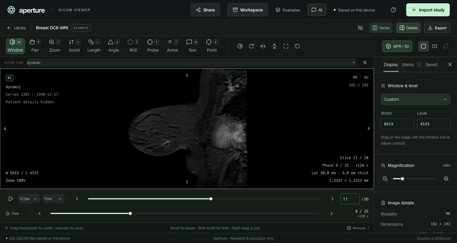
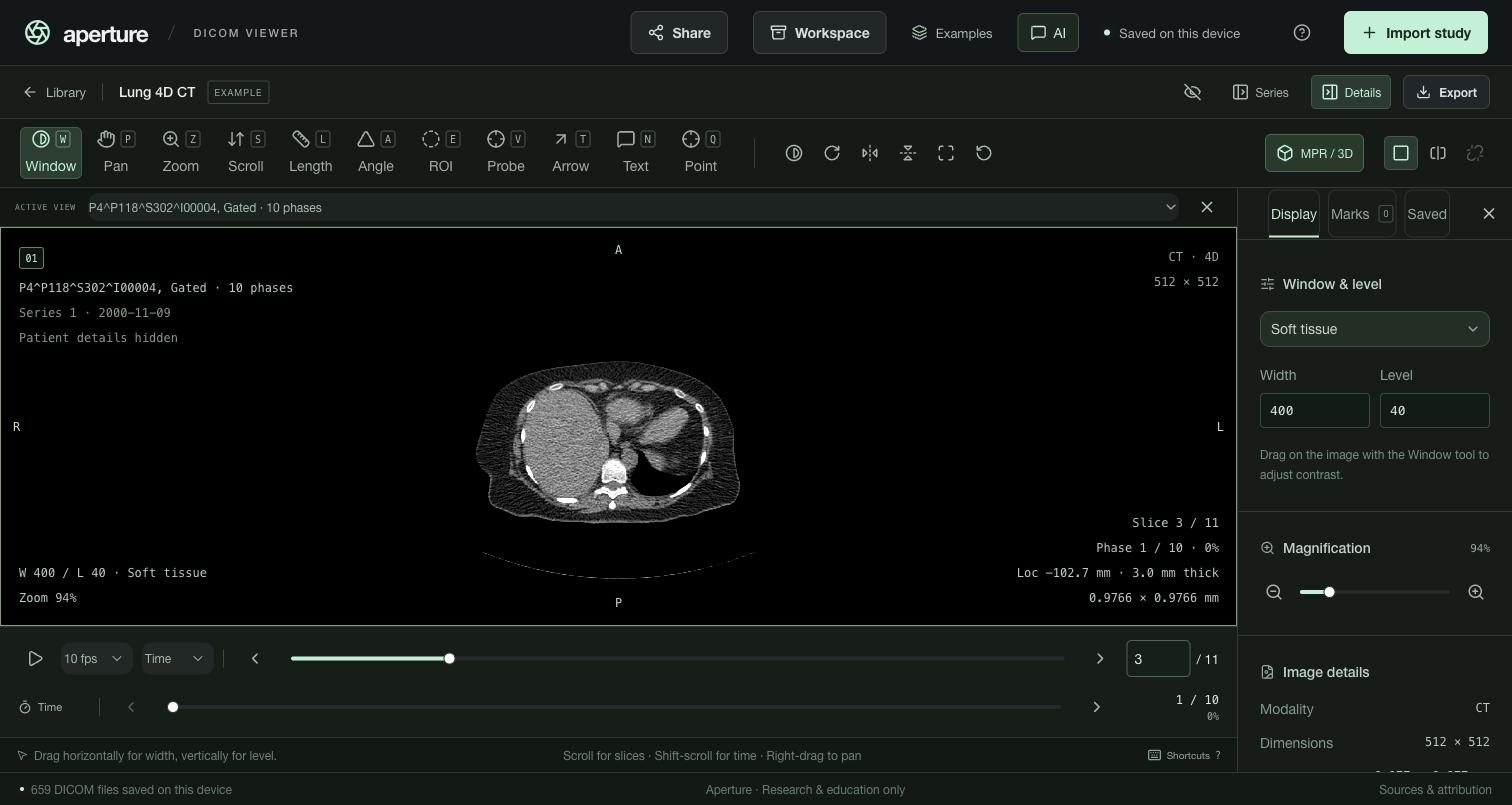
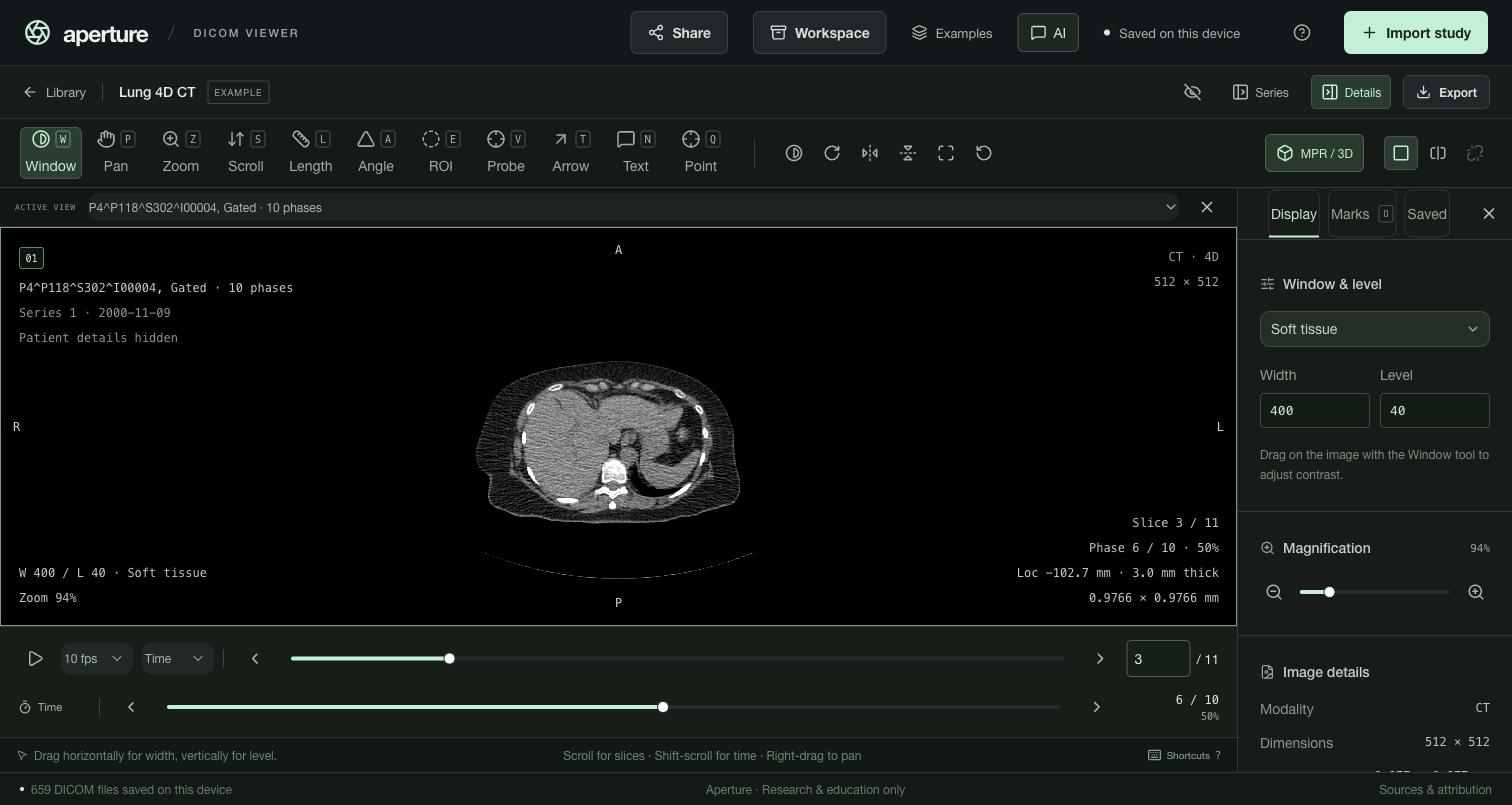
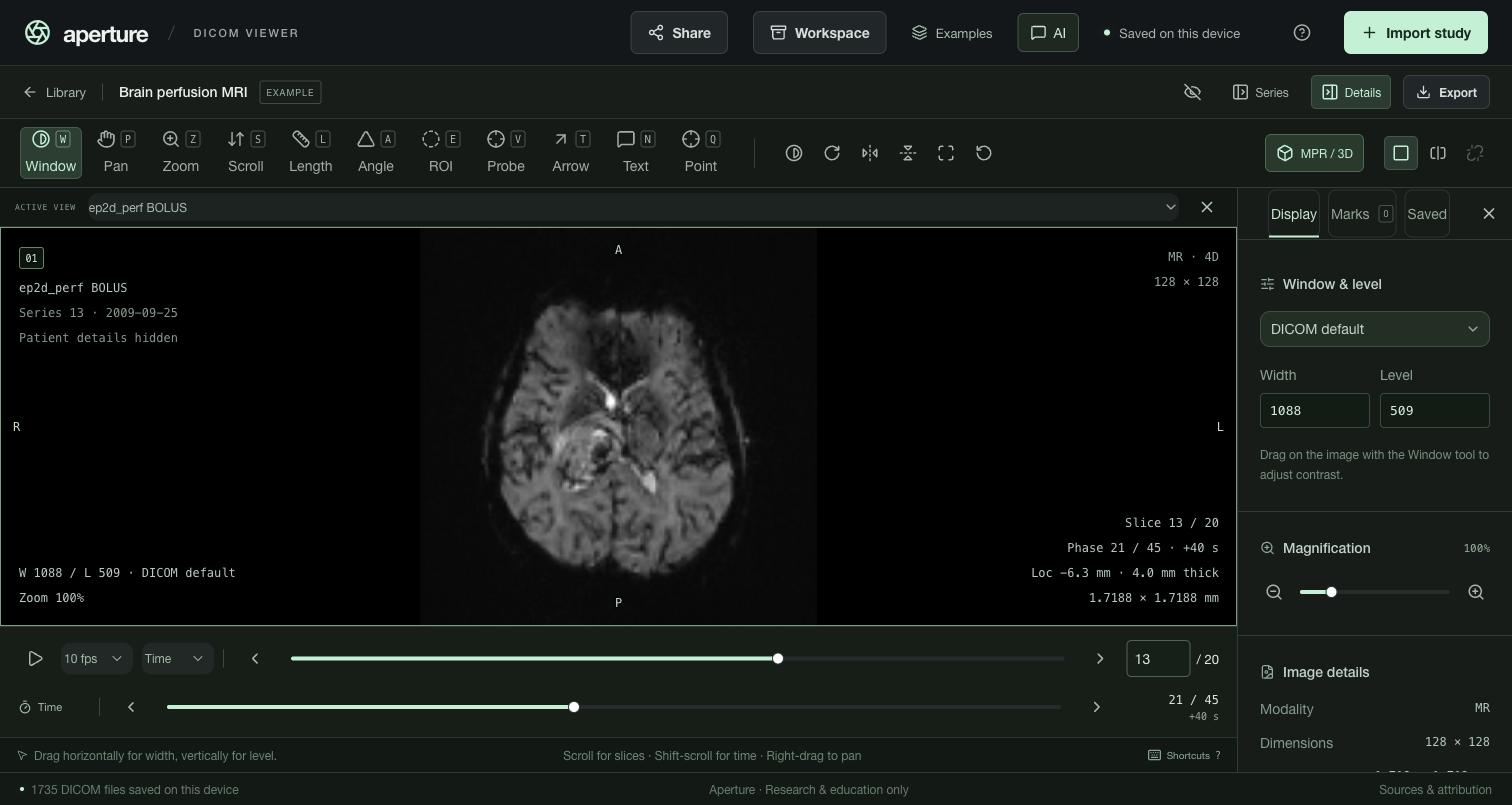
A series is 4D when every slice position repeats and something orders the repeats: cardiac or respiratory phases, or the time points of a contrast study. Aperture reads, in order: temporal position, trigger time, acquisition number, acquisition time, content time, then instance order. Gated CT stored as one series per phase (“Gated, 10.0%”) is joined into one 4D series. 4D series are marked 4D and listed as slices × phases.
- A Time slider sits under the slice slider. The slice slider, arrows and wheel stay within the current phase. , / . or Shift + wheel step through time.
- Cine plays through Time by default; switch it to Slices next to the speed.
- The overlay shows the slice, the phase, and the phase time: milliseconds for cardiac data, seconds since the first phase for contrast series.
- One window holds across all phases, so enhancement or washout reads as real brightness change, not a rescaled image.
4D in MPR / 3D
For a 4D series, MPR / 3D loads every phase into one volume and adds its own Time slider and Play button. The three planes and the 3D rendering change phase in place, keeping crosshairs, zoom and rotation, and the viewer follows to the same phase.
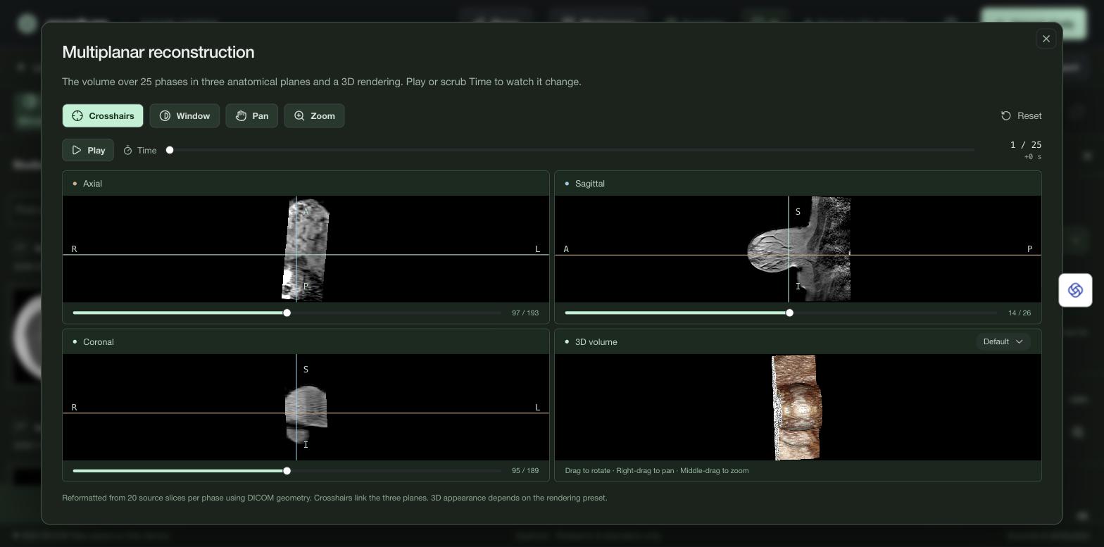
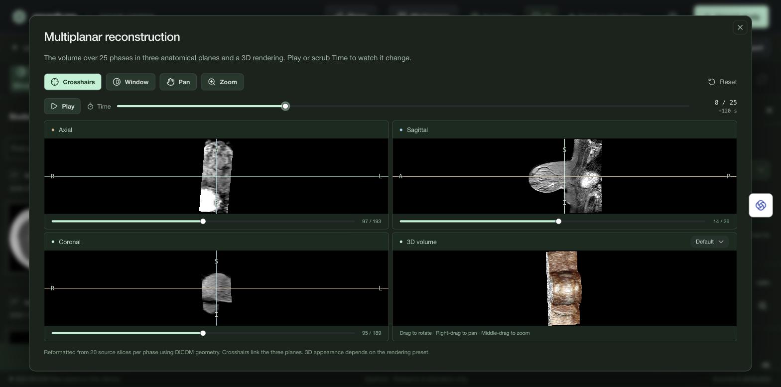
Open Lung 4D CT and press Space to watch it breathe. Then open Breast DCE-MRI, go to slice 11, and step through time with ..
AI assistant
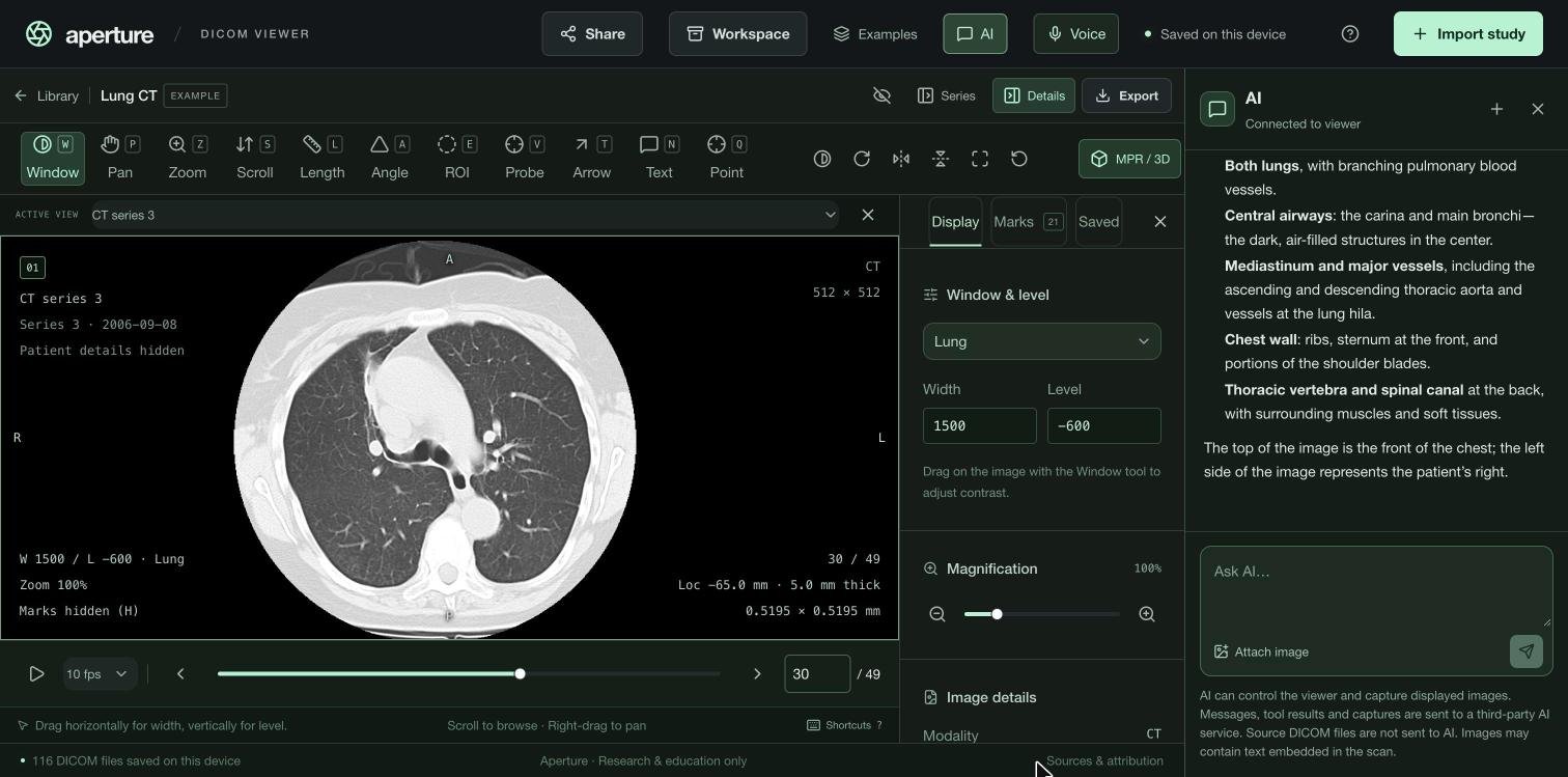
AI opens a chat that can operate the viewer. Plain commands run directly; questions about the image are answered from a capture of the current view. Source DICOM files and patient details are never sent; captures can include text burned into the image. The panel shows each step the assistant took.
In the desktop app, AI features run with your own API key or with Aperture Credits, a monthly subscription that needs no keys.
- Commands: “open the lung CT and use the lung window”, “go to image 20”, “go to phase 8”, “show the other series”.
- Questions: “what is this?”, “which structures are visible on this slice?”.
- Search: “find the largest nodule and mark it”. The assistant steps through the scan, reviews the frames it finds relevant, and places marks you can edit.
- Scan review goes through every image of a series or study in batches, saving observations, marks, key images and notes. Every image answers the same checklist of questions; where images disagree, a stronger model re-checks them before the impression is written from all of the answers. It can pause and resume from its last checkpoint.
- Abdominal CT: “what is abnormal in this abdomen?” screens the whole scan with RADAR. See Abdominal CT screen.
- While AI works, the viewer is locked so a stray click cannot move it. Stop or Take over ends the turn and hands control back.
Abdominal CT screen (RADAR)
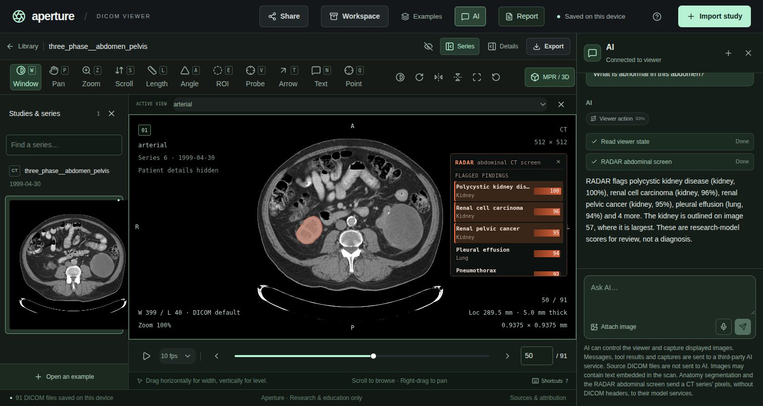
On a contrast-enhanced abdominal CT, the AI assistant can screen the whole scan with RADAR, a research model from Alibaba DAMO Academy (Science, 2026). It was trained on more than 400,000 abdominal CTs and their reports, and scores 146 findings across 18 organs: liver, kidneys, pancreas, spleen, gallbladder, adrenal glands, stomach, duodenum, small and large bowel, esophagus, bladder, aorta, portal vein, heart, lung bases, ribs and sacrum.
- Open the CT series, then open AI.
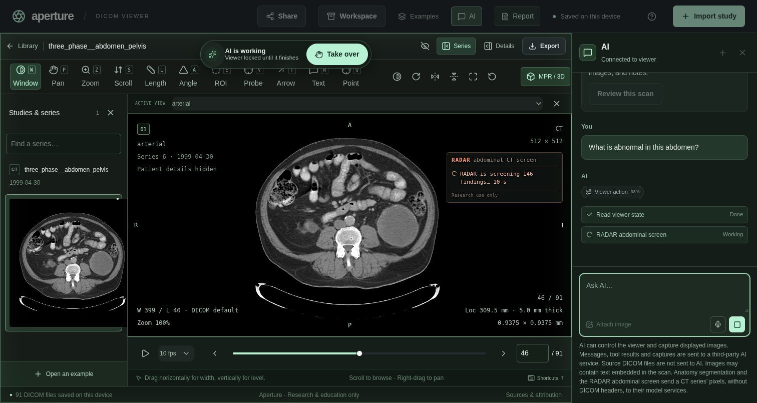
- Ask “What is abnormal in this abdomen?”, “Screen this CT” or “Run RADAR”. A RADAR panel on the image shows the progress, and the viewer is locked until the screen finishes.
- RADAR answers in about 10–20 seconds plus the time to upload the scan (about 10 MB for 90 images). Each series is screened once: the result is saved with the study, so reopening it, reloading the page or asking again is instant.
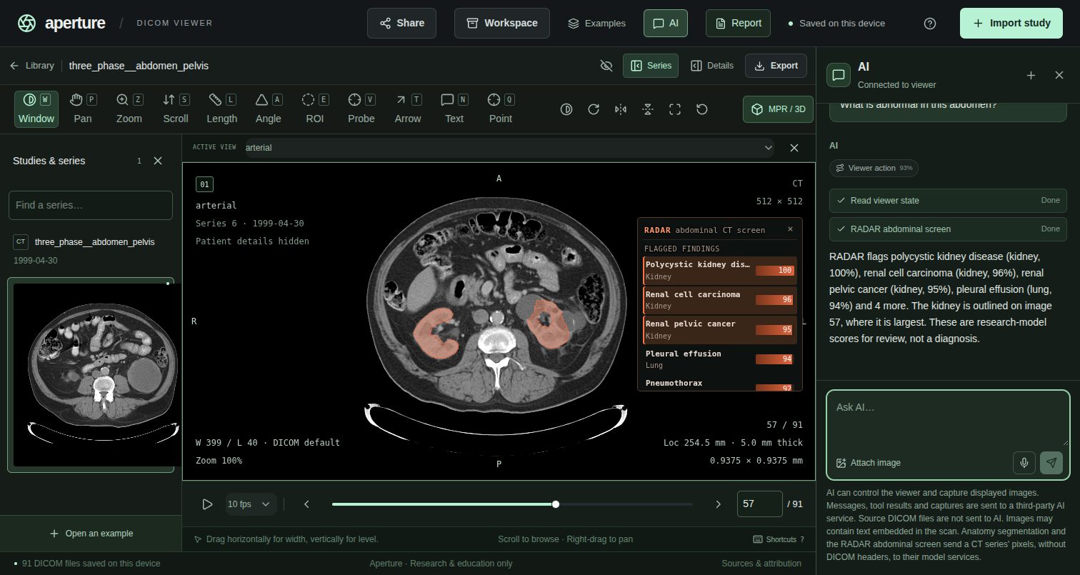
When it finishes, the panel lists the flagged findings, those scoring 50 or more, with the organ each belongs to. When nothing is flagged, it lists the highest scores instead. The organ behind the top finding is outlined in orange, and the view moves to the image where that organ is largest. The chat summarises the same findings.
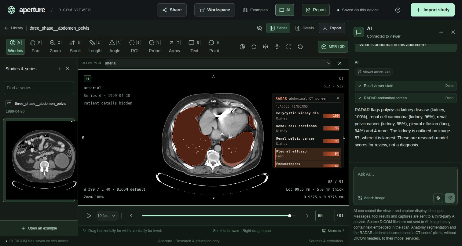
Click any finding to outline its organ and jump to the image where that organ is largest. Click × on the panel to hide it. Asking again brings it back at once from the saved result.
Reading the results
- Scores rank findings; they are not probabilities. RADAR compares the scan with a description of each finding and of its absence. Use the list to decide where to look first.
- RADAR scores whole organs; it does not point to the lesion. The outline is RADAR's map of the organ, not of the abnormality, and it can leave the abnormality out. Step through the outlined images and look for yourself, or ask the assistant to look at the image and mark what it sees.
- Expect false positives. Treat every finding as a suggestion to check, and an empty list as no reason to skip the review.
- It works on contrast-enhanced abdominal CT. Other scans, other body regions and non-contrast CT are not what it was trained on, and its results there are unreliable.
The scan's pixel values and geometry are sent to the RADAR service; DICOM headers and patient details are not. RADAR is a research model licensed for non-commercial use, not a medical device, and its results are not clinically validated.
Download KiTS-00037, arterial series (91 images, 24 MB ZIP) from the C4KC-KiTS collection of The Cancer Imaging Archive (CC BY 3.0). Drop the ZIP on Aperture, open AI and ask “What is abnormal in this abdomen?”. Pathology confirmed a 10 cm clear-cell renal cell carcinoma in the left kidney.
DICOM metadata
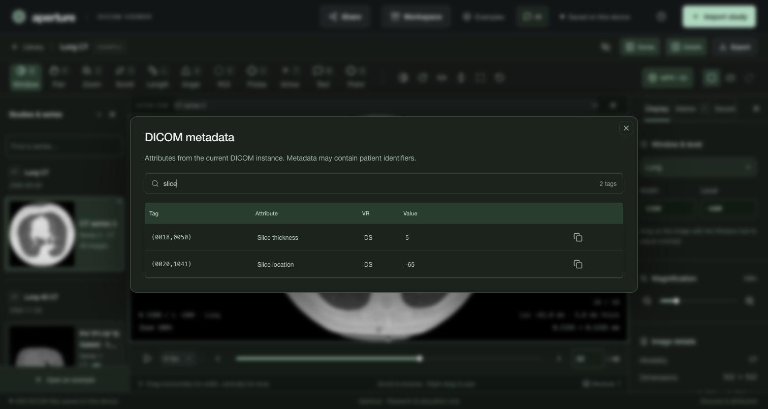
Details summarizes the current image: modality, dimensions, spacing, slice location and thickness, series, and phases for 4D. View DICOM metadata lists every attribute of the current instance, searchable by tag, name or value, with a copy button on each row.
Save, share and export
- Your library, marks, key images, notes and each series' last view are saved to your account and reopen where you left off. Browser Back and Forward move between views, and a view's link reopens the exact image and display for anyone who has the same study.
- Saved keeps named key images and notes attached to exact images.
- Workspace backs up everything to a ZIP and restores it on another device. Share creates an encrypted, expiring link to a workspace.
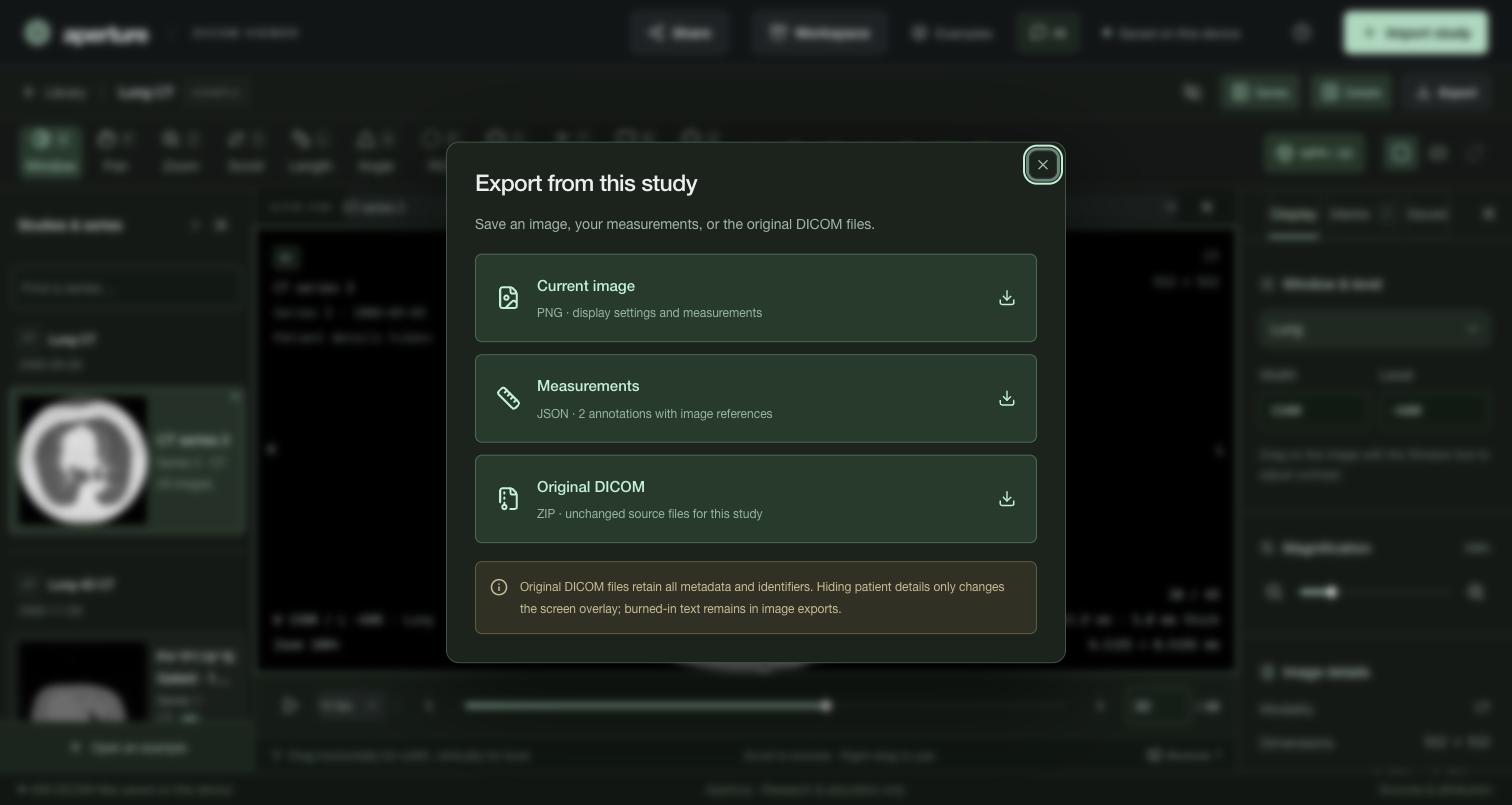
- Export saves the current image as PNG with its display and marks, measurements as JSON, or the study's original DICOM files, unchanged, as a ZIP. Example exports include their license and citation.
Keyboard shortcuts
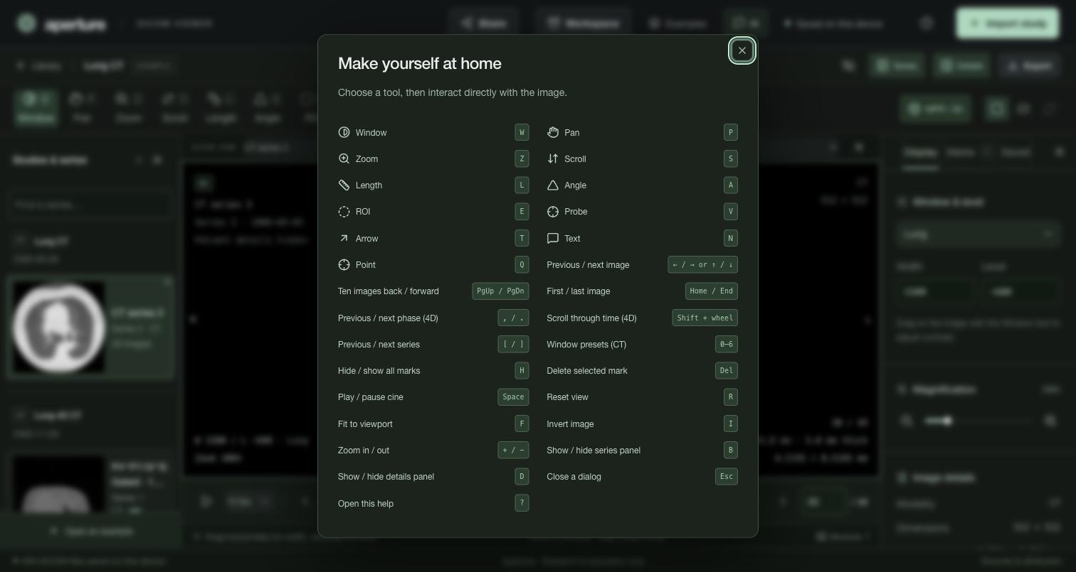
Press ? in the viewer for this list. Shortcuts pause while you type in a field.
Formats, limits and privacy
- Supported: classic CT, MR, CR, DX, ultrasound and secondary capture, including conventional multiframe ultrasound and secondary capture. Uncompressed and JPEG 2000 data are verified on real files; JPEG, JPEG-LS, RLE and HTJ2K decode through Cornerstone's codecs.
- Not supported: enhanced multi-frame CT/MR, segmentations, structured reports, RT objects, whole-slide imaging and video.
- Up to 2,000 files and 512 MiB per import, 256 MiB per file, and 768 MiB across open studies. ZIP archives must not be encrypted, nested or split.
- Hiding patient details changes only the screen overlay. It does not anonymize files, and exports keep all identifiers.
- Measurements and AI output are not clinically validated. Aperture is for research use only. It is not a medical device and must not be used for diagnosis, treatment or any clinical decision.
Example data
All examples are real, deidentified studies from The Cancer Imaging Archive, bundled unchanged. The Lung 4D CT keeps 11 of each phase's 76 slices.
| Example | What it shows | Source | License |
|---|---|---|---|
| Lung CTCT · 3D | Axial chest · soft tissue & lung | RIDER Lung CT | CC BY 4.0 |
| Breast MRIMR · 3D | Axial B0 · magnetic resonance | RIDER Breast MRI | CC BY 3.0 |
| Chest radiographCR | Projection · computed radiography | RIDER Pilot | CC BY 4.0 |
| Brain MRI 3DMR · 3D | 3D · 1 mm isotropic post-contrast T1 · glioblastoma | UPENN-GBM | CC BY 4.0 |
| Breast DCE-MRIMR · 4D | 4D · 20 slices × 25 contrast time points | QIN-BREAST | CC BY 3.0 |
| Lung 4D CTCT · 4D | 4D · lung base over 10 breathing phases | 4D-Lung | CC BY 3.0 |
| Brain perfusion MRIMR · 4D | 4D · 20 slices × 45 time points · contrast bolus | UPENN-GBM | CC BY 4.0 |
| Hip radiographsCR | Orthopedics · AP and frog-leg lateral views | VAREPOP-APOLLO | CC BY 4.0 |
| Shoulder radiographsDX | Orthopedics · internal and external rotation, axial | VAREPOP-APOLLO | CC BY 4.0 |
| Lumbar spine MRIMR | Orthopedics · sagittal T1, T2 and STIR | VAREPOP-APOLLO | CC BY 4.0 |
| Spine CT · metastasesCT · 3D | Orthopedics · whole spine, bone metastases, 3D | Spine-Mets-CT-SEG | CC BY 4.0 |